52 Weeks of Inspiring Illustrations, Week 49: Estienne’s Anatomy (1546)
Charles Estienne’s De dissectione partium corporis humani libri tres (1545) was the third fully illustrated anatomical work ever to be published. Estienne came from a distinguished printing family: his father, Henri, began the family book-trade in the early 16th century and his brother, Robert, improved upon the business by printing elegant Latin and Greek works as well as editions of the New Testament. Although Charles had to take on the family business in 1551, due to Robert fleeing Paris for Geneva (due to persecution for his Protestant beliefs), his heart was in anatomy and medicine. Charles studied in Padua from 1530 to 1534, learning Greek, botany, and natural science. He returned to Paris and began medical study in 1538 under Jacques Dubois (Sylvius), who had taught Andreas Vesalius. He received his medical doctorate in 1542 and was lecturer in anatomy at the Faculty of Medicine from 1544-1547.
It was during this time under Sylvius and his work at the Faculty of Medicine that Estienne began compiling his anatomical atlas. Estienne was key in several important anatomical discoveries including the Hepatic vein of the liver. He gathered his work over several years and by 1539 the manuscript and woodcuts for De dissectione was complete, however as the book was going through the press the publication had to be suspended when Estienne de la Riviére filed a plagiarism suit, demanding credit for having performed the dissections on which the illustrations were based. As a result, publication was delayed for five years. The Latin edition was finally published in 1545, and the French edition (La dissection des parties du corps humain diuisee en trois livres), on display here, probably translated by Estienne himself, was published the following year.
Estienne had hoped to get his work finished and published before Vesalius’s De Corpis Fabrica, surely as a member of a publishing family he was aware that there was only one other fully illustrated anatomical text in existence, that being the work of Jacopo Berengario da Carpi. However, because of the delay his work was published two years after Vesalius’s work, and would ultimately be eclipsed by Vesalius’s scholarship and beautiful illustrations.
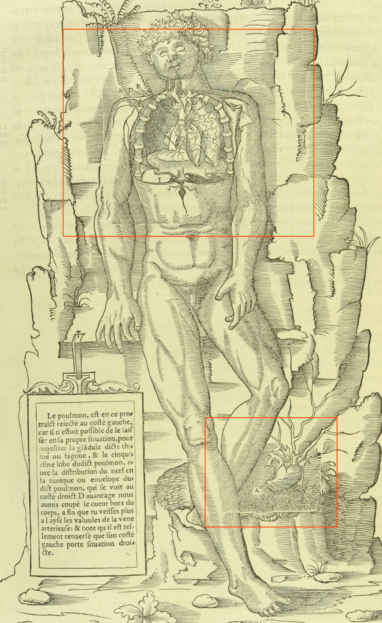
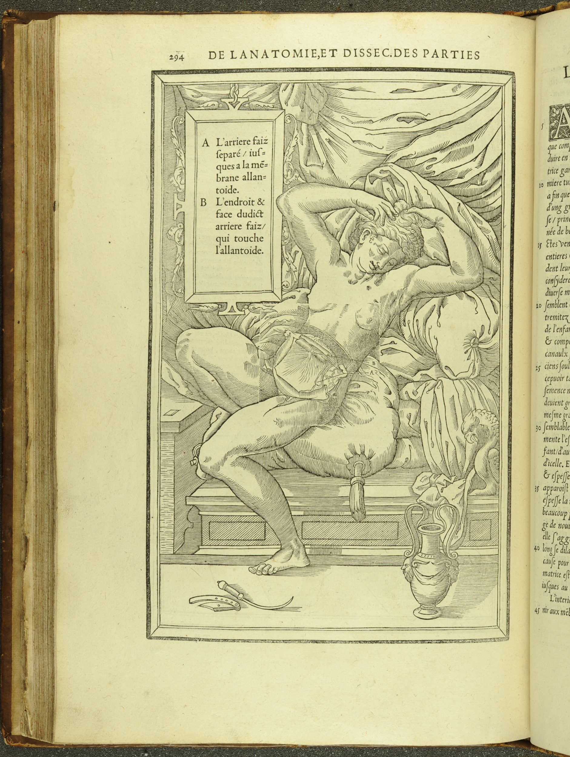
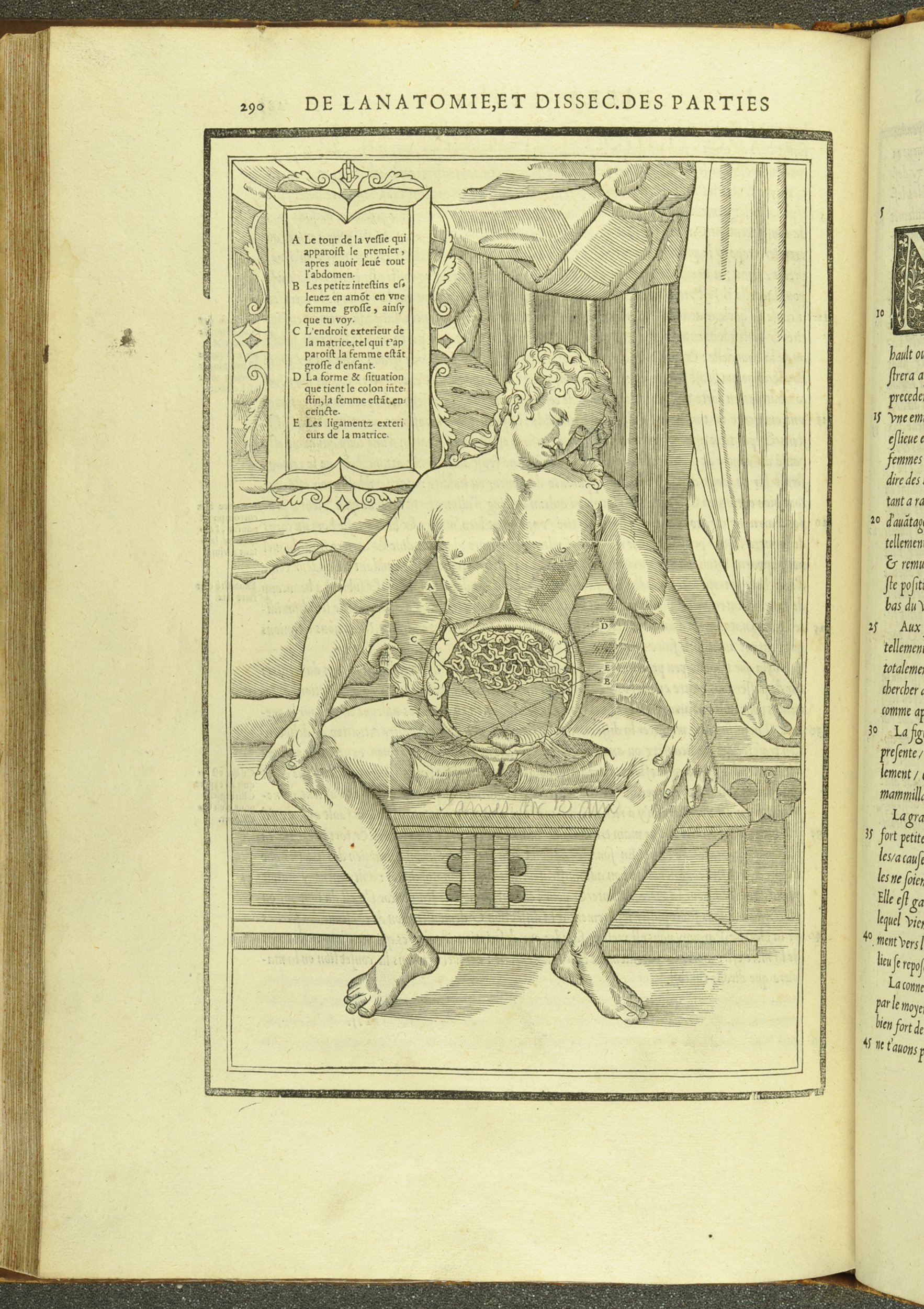
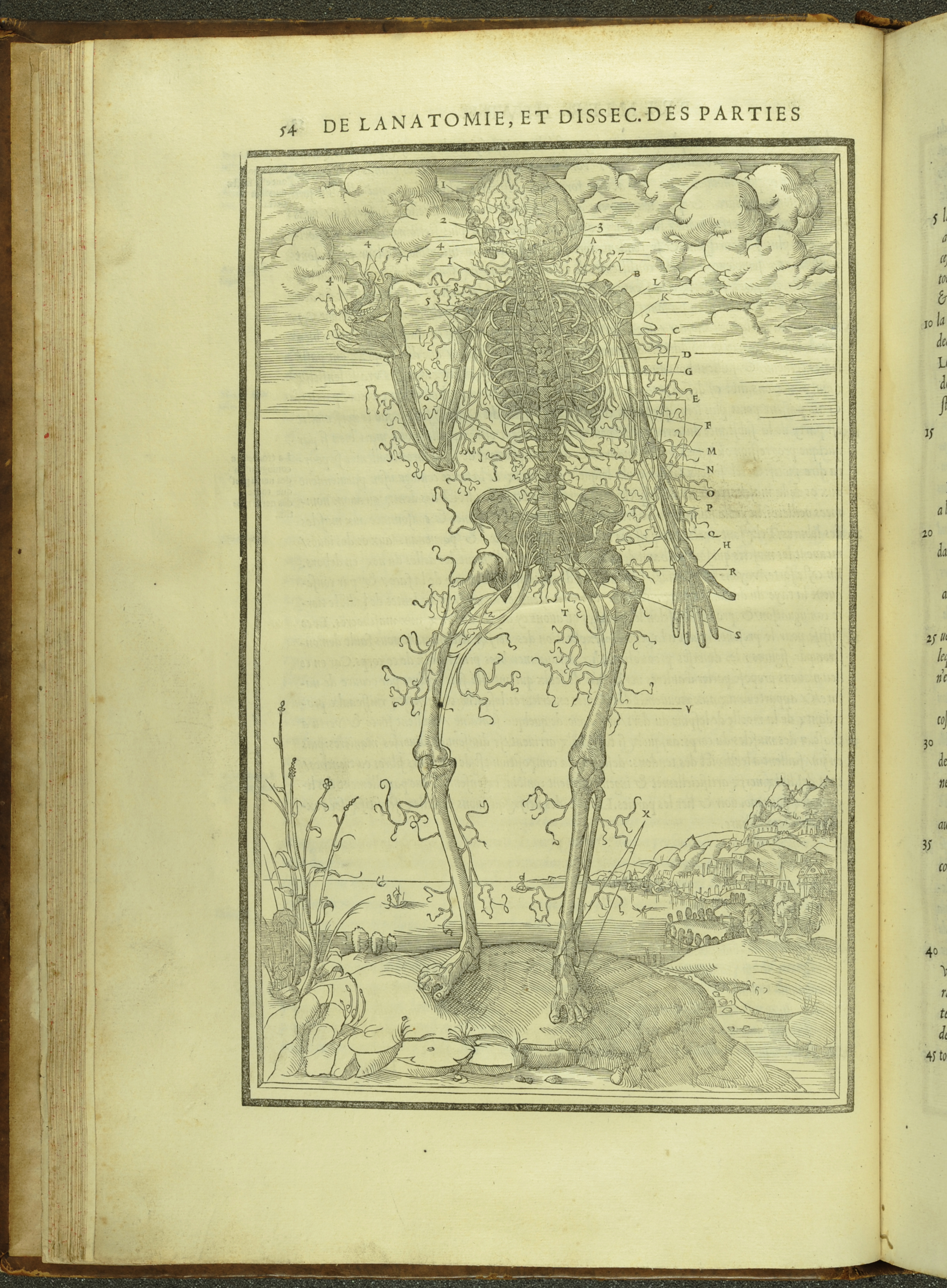
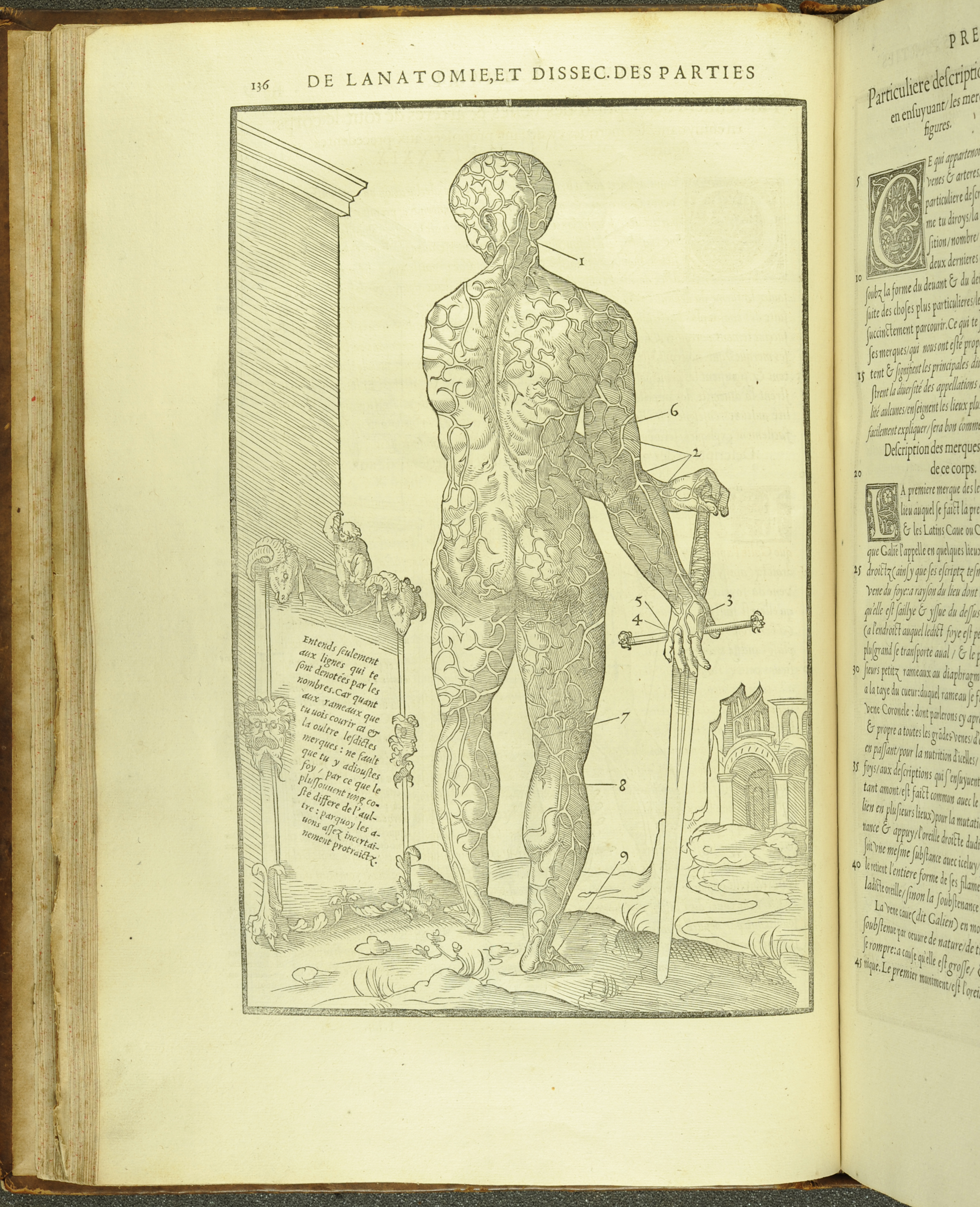
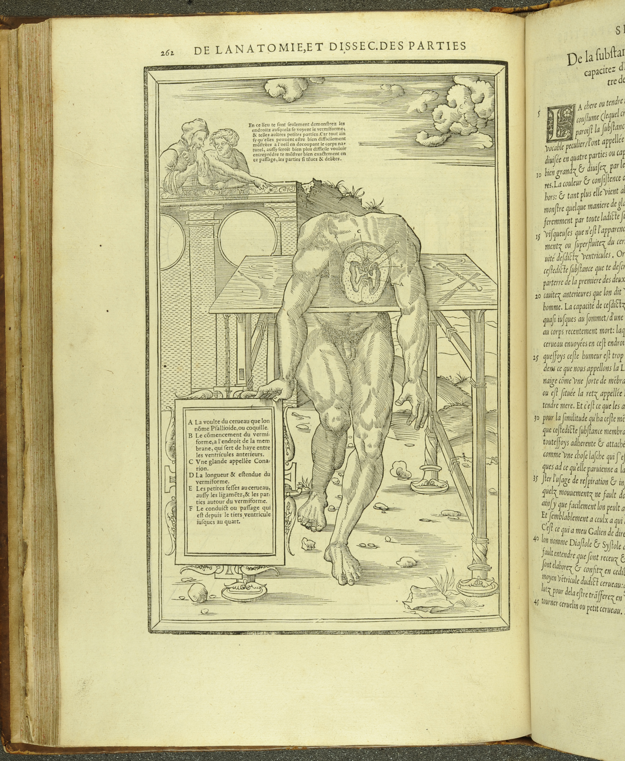
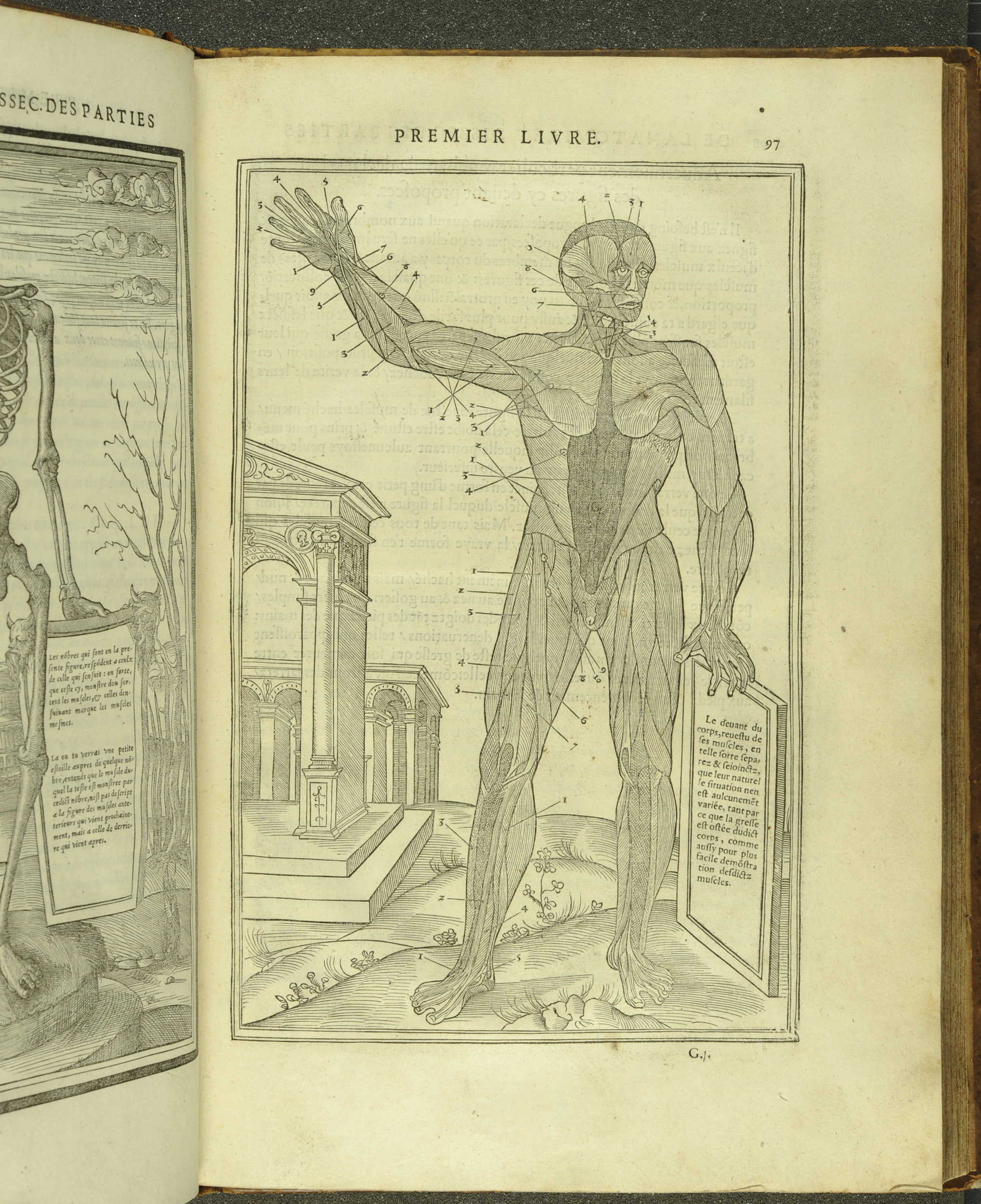
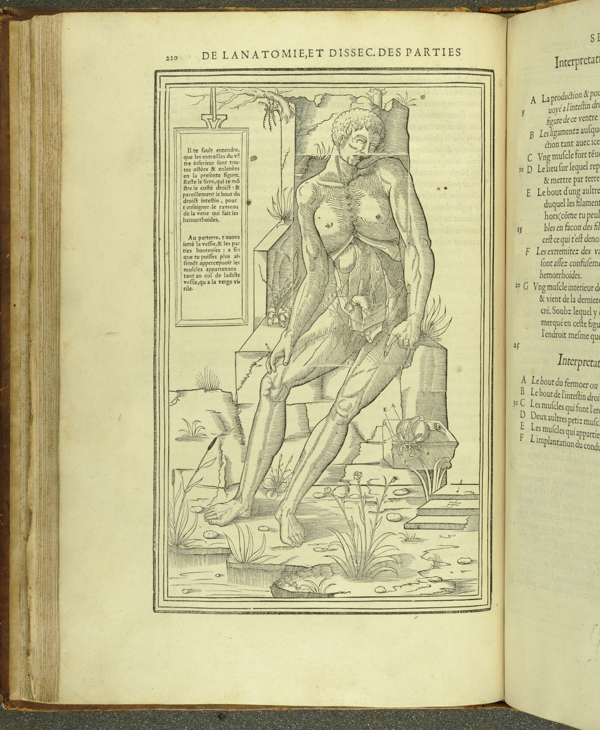
To cut costs, Estienne took some of his illustrations from non-anatomical books, replacing a section of the woodblock with an insert that depicted the body’s interior. This often resulted in uneven matching and muddles the clarity of the anatomical detail being depicted. However, I find some of these anatomical mash-ups quite inspiring, often skeletons or dissected figures are depicted standing outside of some grand, fantastical city. Estienne also used some questionable sources for some of his woodcuts: he illustrated the female reproductive system and gravid uterus by borrowing poses from a series of erotic prints by Jacopo Caraglio that were based on drawings by Perino del Vaga and Rosso Fiorentino.
Whatever the case, this is truly a beautiful book and its illustrations are both artistic and utilitarian. A fully digitised version of this book can be found on Gallica and St Andrews’ copy can be consulted in our Reading Room at any time.
–DG